


Modulation of Erythroid and Immune Cell Markers by Vitamin D and Tinospora cordifolia in infected Mice
Article Sidebar
Views | PDF/EPUB Downloads:
232
/ 101
/ 60
Main Article Content
Abstract
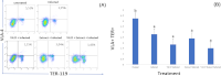
TER-119 is a key marker of mature erythroid cells, which is known for its role in maintaining erythrocyte membrane stability. VLA-4 (CD49d/CD29), an adhesion molecule, facilitates the interaction between immune cells and vascular endothelial cells, especially during inflammation, favoring leukocyte migration. While Vitamin D (VD) and Tinospora cordifolia (TC) are recognized for their immunomodulatory effects, there are limited data regarding their combined effect on erythropoiesis and immune response, particularly about the expression VLA-4 on erithroid cell subset (TER-119) and imunocompeten granulocyte cell subset (Gr-1). This study aimed to evaluate the impact of VD, TC, and their combination on these markers in murine models. The mice were divided into five groups: normal (N), infection control (I), treated with VD (VD, 0.325 μg/kg BW), treated with TC (TC, 100 mg/kg BW), and combination treated (VD+TC). After 28 days of oral treatment, all groups except the normal group received inactivated intraperitoneal injections of E. coli. Flow cytometry revealed that the infection increased the expression of VLA-4 in GR granulocytes (7.03%) compared to the normal group (6.03%). Treatment of VD and TC individually approached expression to normal levels (5.14% and 4.75%, respectively), while their combination further reduced VLA-4 expression to 2.97%. Analysis of VLA-4⁺ TER-119⁺ cells showed the lowest expression in the combination group (1.43%) compared to normal (5.51%), with no statistically significant improvement in erythroid development. These findings suggest that while VD and TC individually modulate the expression of immune markers VLA-4 on erythroid and also on granulocytes, their combination provided additive benefits which was not statistically significant. Further studies are needed to explore the optimal dosage and timing to exploit potential synergistic effects.
Downloads
Article Details
Section

This work is licensed under a Creative Commons Attribution-NonCommercial-NoDerivatives 4.0 International License.
How to Cite
References
1 Zivot A, Lipton JM, Narla A, Blanc L. Erythropoiesis: insights into pathophysiology and treatments in 2017. Mol. Med. 2018: 24:1–15. DOI: https://doi.org/10.1186/s10020-018-0011-z
2 Katiyar S, Shah A, Rahman K, Tripathy NK, Kashyap R, Nityanand S, Chaturvedi CP. Analysis of Immunophenotypic Changes during Ex Vivo Human Erythropoiesis and Its Application in the Study of Normal and Defective Erythropoiesis. Cells 2023:12. https://doi.org/10.3390/cells12091303. DOI: https://doi.org/10.3390/cells12091303
3 Shim YA, Campbell T, Weliwitigoda A, Dosanjh M, Johnson P. Regulation of CD71+TER119+ erythroid progenitor cells by CD45. Exp. Hematol. 2020: 86: 53-66. e1. https://doi.org/10.1016/j.exphem.2020.05.005. DOI: https://doi.org/10.1016/j.exphem.2020.05.005
4 Härzschel A, Zucchetto A, Gattei V, Hartmann TN. VLA-4 Expression and Activation in B Cell Malignancies: Functional and Clinical Aspects. Int. J. of Mol Sci. 2020:21. https://doi.org/10.3390/ijms21062206. DOI: https://doi.org/10.3390/ijms21062206
5 Berrazouane S, Doucet A, Boisvert M, Barabé F, Aoudjit F. Vla-4 induces chemoresistance of t cell acute lymphoblastic leukemia cells via pyk2-mediated drug efflux. Cancers. 2021:13. https://doi.org/10.3390/cancers13143512. DOI: https://doi.org/10.3390/cancers13143512
6 Bishop EL, Ismailova A, Dimeloe S, Hewison M, White JH. Vitamin D and Immune Regulation: Antibacterial, Antiviral, Anti-Inflammatory. JBMR Plus 2021:5:1–23. https://doi.org/10.1002/jbm4.10405. DOI: https://doi.org/10.1002/jbm4.10405
7 Arora J, Wang J, Weaver V, Zhang Y, Cantorna MT. Novel insight into the role of the vitamin D receptor in the development and function of the immune system. J Steroid Biochem Mol Biol 2023:291:1–19. https://doi.org/10.1016/j.jsbmb.2022.106084.Novel.
8 Gao H, Zhou H, Zhang Z, Gao J, Li J, Li X. Vitamin D3 alleviates inflammation in ulcerative colitis by activating the VDR-NLRP6 signaling pathway. Front. Immunol. 2023: 14:1–12. https://doi.org/10.3389/fimmu.2023.1135930. DOI: https://doi.org/10.3389/fimmu.2023.1135930
9 Fletcher J, Bishop EL, Harrison SR, Swift A, Cooper SC, Dimeloe SK, Raza K, Hewison M. Autoimmune disease and interconnections with vitamin D. Endo Connect. 2022:11. https://doi.org/10.1530/EC-21-0554. DOI: https://doi.org/10.1530/EC-21-0554
10 Ahsan R, Mishra A, Badar B, Owais M, Mishra V. Therapeutic Application, Phytoactives and Pharmacology of Tinospora cordifolia: An Evocative Review. Chin J of Int Med. 2023:29:549–55. https://doi.org/10.1007/s11655-023-3733-2. DOI: https://doi.org/10.1007/s11655-023-3733-2
11 Rahman AA, Rahman S, Jahan G, Sarder M. Phytochemical and Pharmacological Investigations of Tinospora Cordifolia. Khul Univ Stud. 2022. DOI: https://doi.org/10.53808/KUS.2006.7.1.0533-L
12 Gupta PK, Rajan MGR, Kulkarni S. Activation of murine macrophages by G1-4A, a polysaccharide from Tinospora cordifolia, in TLR4/MyD88 dependent manner. Int Immun. 2017:50:168–77. https://doi.org/10.1016/j.intimp.2017.06.025. DOI: https://doi.org/10.1016/j.intimp.2017.06.025
13 Yates CR, Bruno EJ, Yates MED. Tinospora Cordifolia: A review of its immunomodulatory properties. J Diet Suppl 2022:19:271–85. https://doi.org/10.1080/19390211.2021.1873214. DOI: https://doi.org/10.1080/19390211.2021.1873214
14 Philip S, Tom G, Balakrishnan Nair P, Sundaram S, Velikkakathu Vasumathy A. Tinospora cordifolia chloroform extract inhibits LPS-induced inflammation via NF-κB inactivation in THP-1cells and improves survival in sepsis. BMC Comp Med Ther. 2021:21:1–13. https://doi.org/10.1186/s12906-021-03244-y. DOI: https://doi.org/10.1186/s12906-021-03244-y
15 Jamil AS, Widyarti S, Setiawan M, Rifa’i M. Bioactivity of Vitamin D and Tinospora cordifolia Extract in Increasing Cathelicidin Synthesis and Regulating TNF Alpha Production in CD66a Cells. Res Res Biomed. 2025:11:111–9. https://doi.org/10.18413/2658-6533-2025-11-1-0-6. DOI: https://doi.org/10.18413/2658-6533-2025-11-1-0-6
16 Zahiruddin S, Parveen A, Khan W, Ibrahim M, Want MY, Parveen R, Ahmad S. Metabolomic Profiling and Immunomodulatory Activity of a Polyherbal Combination in Cyclophosphamide-Induced Immunosuppressed Mice. Front Pharm. 2022:12:1–15. https://doi.org/10.3389/fphar.2021.647244. DOI: https://doi.org/10.3389/fphar.2021.647244
17 Wuryandari MRE, Atho’illah MF, Laili RD, Fatmawati S, Widodo N, Widjajanto E, Rifa’i M. Lactobacillus plantarum FNCC 0137 fermented red Moringa oleifera exhibits protective effects in mice challenged with Salmonella typhi via TLR3/TLR4 inhibition and down-regulation of proinflammatory cytokines. J Ayurveda Integr Med 2022:13:100531. https://doi.org/10.1016/j.jaim.2021.10.003. DOI: https://doi.org/10.1016/j.jaim.2021.10.003
18 Atho’illah MF, Safitri YD, Nur’aini FD, Widyarti S, Tsuboi H, Rifa’i M. Elicited soybean extract attenuates proinflammatory cytokines expression by modulating TLR3/TLR4 activation in high−fat, high−fructose diet mice. J Ayurveda Integr Med 2021:12:43–51. https://doi.org/10.1016/j.jaim.2021.01.003. DOI: https://doi.org/10.1016/j.jaim.2021.01.003
19 Safitri YD, Atho’illah MF, Nur’aini FD, Widyarti S, Rifa’i M. The Effects of Elicited Soybean (Glycine max) Extract on. Jor J Biol Sci. 2018:11:241–246.
20 Lestari SR, Christina YI, Atho’illah MF, Rifa’i M. Single-bulb garlic oil regulates toll-like receptors and Nrf2 cross-talk and IL-17 production in mice fed with high-fat diet. Saudi Journal of Biological Sciences 2021:28:6515–6522. https://doi.org/10.1016/j.sjbs.2021.07.021. DOI: https://doi.org/10.1016/j.sjbs.2021.07.021
21 Verdegaal EME, Beekhuizen H, Blokland I, Van Furth R. Increased adhesion of human monocytes to IL-4-stimulated human venous endothelial cells via CD11/CD18, and very late antigen-4 (VLA-4)/vascular cell adhesion molecule-1 (VCAM-1)-dependent mechanisms. Clinical and Experimental Immunology 1993:93:292–298. https://doi.org/10.1111/j.1365-2249.1993.tb07982.x. DOI: https://doi.org/10.1111/j.1365-2249.1993.tb07982.x
22 Jung O, Beauvais DM, Adams KM, Rapraeger AC. VLA-4 phosphorylation during tumor and immune cell migration relies on its coupling to VEGFR2 and CXCR4 by syndecan-1. Journal of Cell Science 2019:132. https://doi.org/10.1242/jcs.232645. DOI: https://doi.org/10.1242/jcs.232645
23 Schröder-Heurich B, von Hardenberg S, Brodowski L, Kipke B, von Meyer N, Borns K, Kaisenberg CS, Brinkmann H, Claus P, von Versen-Höynck F. Vitamin D improves endothelial barrier integrity and counteracts inflammatory effects on endothelial progenitor cells. FASEB Journal 2019:33:9142–9153. https://doi.org/10.1096/fj.201802750RR. DOI: https://doi.org/10.1096/fj.201802750RR
24 Kristianti MTF, Goenawan H, Achadiyani A, Sylviana N, Lesmana R. The Potential Role of Vitamin D Administration in The Skin Aging Process Through the Inflammatory Pathway: A Systematic Review. Trop J Nat Prod Res. 2023:7:2675–2681. https://doi.org/10.26538/tjnpr/v7i4.1. DOI: https://doi.org/10.26538/tjnpr/v7i4.1
25 Mahlknecht U, Schönbein C. Histone deacetylase inhibitor treatment downregulates VLA-4 adhesion in hematopoietic stem cells and acute myeloid leukemia blast cells. Haematologica 2008:93:443–446. https://doi.org/10.3324/haematol.11796. DOI: https://doi.org/10.3324/haematol.11796
26 Nandan A, Sharma V, Banerjee P, Sadasivam K, Venkatesan S, Prasher B. Deciphering the mechanism of Tinospora cordifolia extract on Th17 cells through in-depth transcriptomic profiling and in silico analysis. Front Pharm. 2023:13:1–18. https://doi.org/10.3389/fphar.2022.1056677. DOI: https://doi.org/10.3389/fphar.2022.1056677
27 Philip S, Tom G, Vasumathi A V. Evaluation of the anti-inflammatory activity of Tinospora cordifolia (Willd.) Miers chloroform extract – a preclinical study. J Pharm Pharmacol. 2018:70:1113–1125. https://doi.org/10.1111/jphp.12932. DOI: https://doi.org/10.1111/jphp.12932
28 Avemaria F, Gur-Cohen S, Avci S, Lapidot T. VLA-4 Affinity Assay for Murine Bone Marrow-derived Hematopoietic Stem Cells. Bio-Protocol 2017:7:1–9. https://doi.org/10.21769/bioprotoc.2134. DOI: https://doi.org/10.21769/BioProtoc.2134
29 Bousounis P, Bergo V, Trompouki E. Inflammation, Aging and Hematopoiesis: A Complex Relationship. Cells 2021:1–15. DOI: https://doi.org/10.3390/cells10061386
30 Najafi S, Ghanavat M, Shahrabi S, Gatavizadeh Z, Saki N. The effect of inflammatory factors and their inhibitors on the hematopoietic stem cells fate. Cell Biol Int. 2021:45:900–12. https://doi.org/10.1002/cbin.11545. DOI: https://doi.org/10.1002/cbin.11545
31 Paulson RF, Ruan B, Hao S, Chen Y. Stress Erythropoiesis is a Key Inflammatory Response. Cells 2020;9:634. https://doi.org/10.3390/cells9030634. DOI: https://doi.org/10.3390/cells9030634
32 Yang C, Suda T. Microenvironmental dynamics in steady-state and stress erythropoiesis. Blood Sci. 2025;7:e00219. https://doi.org/10.1097/BS9.0000000000000219. DOI: https://doi.org/10.1097/BS9.0000000000000219
33 Wang L, Degheidy H, Abbasi F, Mostowski H, Marti G, Bauer S, Hoffman RA, Gaigalas AK. Quantitative flow cytometry measurements in antibodies bound per cell based on a CD4 reference. Cur Prot Cyto. 2016:2016:1.29.1-1.29.14. https://doi.org/10.1002/0471142956.cy0129s75. DOI: https://doi.org/10.1002/0471142956.cy0129s75
34 Pitoiset F, Cassard L, El Soufi K, Boselli L, Grivel J, Roux A, Klatzmann D, Chaput N, Rosenzwajg M. Deep phenotyping of immune cell populations by optimized and standardized flow cytometry analyses. Cyto Part A. 2018:93:793–802. https://doi.org/10.1002/cyto.a.23570. DOI: https://doi.org/10.1002/cyto.a.23570