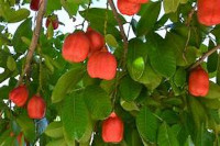
Toxicological Evaluation of the Aqueous Leaf Extract of Blighia Sapida K.D. Koenig (Sapindaceae) in Rodents http://www.doi.org/10.26538/tjnpr/v7i12.48
Main Article Content
Abstract
The leaf of Blighia sapida K.D. Koenig (Sapindaceae) has been used traditionally to treat several conditions, such as diabetes mellitus and hypertension. However, there is still limited knowledge of its toxicity and safety. The study was aimed at investigating the sub-acute toxicological profile of the aqueous leaf extract of Blighia sapida in rodents. The leaf extract was obtained by cold maceration for 72 h. A 21-day sub-acute toxicity test was performed using doses of 500, 1000, and 2000 mg/kg orally. In vitro, antioxidant tests were performed using standard methods 2, 2-diphenyl-2-picryl-hydrazyl, ferric reducing antioxidant power, 2, 2'-azino-bis (3-ethylbenzthiazoline-6-sulphonate) and hydroxyl tests. Biochemical, hematological and histological studies were performed. The leaf extract demonstrated antioxidant activity. The spleen was significantly increased at 2000 mg/kg compared to the control. Marked increases in hemoglobin levels at all the doses, hematocrit levels at 500 mg/kg and platelet counts at 1000 mg/kg relative to the control were observed. Cholesterol and triglyceride levels were significantly decreased at 500 and 1000 mg/kg in relation to the control. Urea concentrations at 1000 and 2000 mg/kg were significantly increased relative to the control. A significant increase was observed in albumin levels at 500 and 2000 mg/kg compared to the control. Alkaline phosphatase was reduced at 2000 mg/kg compared to the control. Most organs showed normal histological features at 500 mg/kg. This study shows that repeated exposure of the extract to rats may be toxic at doses above 500 mg/kg and may be unsafe for traditional users at higher doses.
Downloads
Article Details

This work is licensed under a Creative Commons Attribution-NonCommercial-NoDerivatives 4.0 International License.
References
Setzer RW, Kimmel CA. Use of NOAEL, benchmark dose, and other models for human risk assessment of hormonally active substances. Pure Appl Chem. 2003;75(11–12):2151–8.
Wilson NH, Hardisty JF HJ. Short-term, subchronic, and chronic toxicology studies. Principles and Methods of Toxicology. fourth edi. Francis T and, editor. Philadelphia: Elsevier; 2001. 917-57. p.
Anadón A, Martínez MA, Castellano V, Martínez-Larrañaga MR. The role of in vitro methods as alternatives to animals in toxicity testing. Expert Opin Drug Metab Toxicol. 2014;10(1):67–79.
Onuekwusi EC, Akanya HO, Evans EC. Phytochemical Constituents of Seeds of Ripe and Unripe Blighia sapida (K. Koenig) and Physicochemical Properties of the Seed Oil [Internet]. Vol. 3, Int. J Pharmaceutical Sci. Invent. ISSN. Online; 2014 [cited 2020 Dec 1]. Available from: www.ijpsi.org
Abolaji AO, Adebayo HA, Odesanmi OS. Nutritional Qualities of Three Medicinal Plant Parts (Xylopia aethiopica, Blighia sapida and Parinari polyandra) commonly used by Pregnant Women in the Western Part of Nigeria. 2007;
Marles RJ FN. Antidiabetic plants and their active constituents. Phytomedicine. 1995;2:137–189.
Okogun J. I. The chemistry of Nigerian medicinal plants. 1996;10:31–5.
Etukudo I. Ethnobotany. Conventional and Traditional Uses of Plants. Lippincott Williams & Wilkins; 2003.
Olayinka JN, Ozolua RI, Akhigbemen AM. Phytochemical screening of aqueous leaf extract of Blighia sapida KD Koenig (Sapindaceae) and its analgesic property in mice. J Ethnopharmacol. 2021;273:113977.
Parkinson AA. Phytochemical analysis of ackee (Blighia sapida) pods. City University of New York; 2007.
Mazzola EP, Parkinson A, Kennelly EJ, Coxon B, Einbond LS, Freedberg DI. Utility of coupled-HSQC experiments in the intact structural elucidation of three complex saponins from Blighia sapida. Carbohydr Res. 2011 May 1;346(6):759–68.
John-Dewole OO, Agunbiade SO, Alao OO, Arojojoye OA. Phytochemical and antimicrobial studies of extract of the fruit of Xylopia aethiopica for medicinal importance. E3 J Biotechnol Pharm Res. 2012;3(6):118–22.
Oloyede OB, Ajiboye TO, Abdussalam AF, Adeleye AO. Blighia sapida leaves halt elevated blood glucose, dyslipidemia and oxidative stress in alloxan-induced diabetic rats. J Ethnopharmacol. 2014;157:309–19.
Von Holt C. Methylenecyclopropaneacetic acid, a metabolite of hypoglycin. Biochim Biophys Acta (BBA)-Lipids Lipid Metab. 1966;125(1):1–10.
Moya J. Ackee (Blighia sapida) poisoning in the Northern Province, Haiti, 2001. Epidemiol Bull. 2001;22(2):8–9.
Ajayi AM, Ayodele EO, Ben-Azu B, Aderibigbe AO, Umukoro S. Evaluation of neurotoxicity and hepatotoxicity effects of acute and sub-acute oral administration of unripe ackee (Blighia sapida) fruit extract. Toxicol Reports. 2019;6:656–65.
Tanaka K, Ikeda Y. Hypoglycin and Jamaican vomiting sickness. Prog Clin Biol Res. 1990;321:167–84.
Barceloux DG. Akee fruit and Jamaican vomiting sickness (Blighia sapida Köenig). Dis DM. 2009;55(6):318–26.
Chang S-T, Wu J-H, Wang S-Y, Kang P-L, Yang N-S, Shyur L-F. Antioxidant activity of extracts from Acacia confusa bark and heartwood. J Agric Food Chem. 2001;49(7):3420–4.
Benzie IFF, Strain JJ. [2] Ferric reducing/antioxidant power assay: direct measure of total antioxidant activity of biological fluids and modified version for simultaneous measurement of total antioxidant power and ascorbic acid concentration. In: Methods in enzymology. Elsevier; 1999. p. 15–27.
Kunchandy E, Rao MNA. Oxygen radical scavenging activity of curcumin. Int J Pharm. 1990;58(3):237–40.
Stratil P, Klejdus B, Kubáň V. Determination of total content of phenolic compounds and their antioxidant activity in vegetables evaluation of spectrophotometric methods. J Agric Food Chem. 2006;54(3):607–16.
Richmond W. Preparation and properties of a cholesterol oxidase from Nocardia sp. and its application to the enzymatic assay of total cholesterol in serum. Clin Chem. 1973;19(12):1350–6.
Allain CC, Poon LS, Chan CSG, Richmond W, Fu PC. Enzymatic determination of total serum cholesterol. Clin Chem. 1974;20(4):470–5.
Lopes-Virella MF, Stone P, Ellis S, Colwell JA. Cholesterol determination in high-density lipoproteins separated by three different methods. Clin Chem. 1977;23(5):882–4.
Fossati P, Prencipe L. Serum triglycerides determined colorimetrically with an enzyme that produces hydrogen peroxide. Clin Chem. 1982;28(10):2077–80.
Wieland H, Seidel D. A simple specific method for precipitation of low density lipoproteins. J Lipid Res. 1983;24(7):904–9.
Fawcett J, Scott J. A rapid and precise method for the determination of urea. J Clin Pathol. 1960;13(2):156–9.
Brod J, Sirota JH. The renal clearance of endogenous “creatinine” in man. J Clin Invest. 1948;27(5):645–54.
Onunogbo CC, Ohaeri OC, Eleazu CO. Effect of mistletoe (Viscum album) extract on the blood glucose, liver enzymes and electrolyte balance in alloxan induced diabetic rats. Am J Biochem Mol Biol. 2013;3:143–50.
Walter, K, Schutt C. Acid and alkaline phosphatase in serum (two point method). Bergmeyer HU, editor. Vol. 2, Methods of Enzymatic Analysis. Weinheim,: Verlag Chemie & Academic Press,; 1974.
Reitman S, Frankel S. A colorimetric method for the determination of serum glutamic oxalacetic and glutamic pyruvic transaminases. Am J Clin Pathol. 1957;28(1):56–63.
Tietz NW, Finley PR, Pruden EL. Clinical guide to laboratory tests. Vol. 624. WB Saunders company Philadelphia; 1995.
Jendrassik L, Grof P. Colorimetric method of determination of bilirubin. Biochem z. 1938;297(81):e2.
Blanckaert N. Analysis of bilirubin and bilirubin mono-and di-conjugates. Determination of their relative amounts in biological samples. Biochem J. 1980;185(1):115–28.
Doumas BT, Watson WA, Biggs HG. Albumin standards and the measurement of serum albumin with bromcresol green. Clin Chim acta. 1971;31(1):87–96.
Grant GH, Silverman LM, Chistenson RH. Amino acids and protein in: Tietz NW, ed. Fundamental of clinical chemistry. 3 rd. Philadelphia, WB Saunders Company; 1987.
Adefemi OA, Elujoba AA, Odesanmi WO. Evaluation of the toxicity potential of Cassia podocarpa with reference to official Senna. West Afr J Pharmacol Drug Res. 1988;8:41–8.
Nirogi R, Goyal VK, Jana S, Pandey SK, Gothi A. What suits best for organ weight analysis: review of relationship between organ weight and body/brain weight for rodent toxicity studies. Int J Pharm Sci Res. 2014;5(4):1525–32.
Kern WF. PDQ hematology. PMPH-USA; 2002.
Gotoh S, Hata J, Ninomiya T, Hirakawa Y, Nagata M, Mukai N, et al. Hematocrit and the risk of cardiovascular disease in a Japanese community: The Hisayama Study. Atherosclerosis. 2015;242(1):199–204.
Kishimoto S, Maruhashi T, Kajikawa M, Matsui S, Hashimoto H, Takaeko Y, et al. Hematocrit, hemoglobin and red blood cells are associated with vascular function and vascular structure in men. Sci Rep. 2020;10(1):1–9.
Radaelli F, Colombi M, Calori R, Zilioli VR, Bramanti S, Iurlo A, et al. Analysis of risk factors predicting thrombotic and/or haemorrhagic complications in 306 patients with essential thrombocythemia. Hematol Oncol. 2007;25(3):115–20.
Xu QQ, Xu YJ, Yang C, Tang Y, Li L, Cai H Bin, et al. Sodium Tanshinone IIA Sulfonate Attenuates Scopolamine-Induced Cognitive Dysfunctions via Improving Cholinergic System. Biomed Res Int. 2016;2016.
LaRegina MC, Sharp PE. The Laboratory Rat: A Volume in the Laboratory Animal Pocket Reference Series. CRC Press USA Hal. 1998;1(9):17–8.
Oakenfull DG, Fenwick DE. Adsorption of bile salts from aqueous solution by plant fibre and cholestyramine. Br J Nutr. 1978;40(2):299–309.
Wan C, Wong CN, Pin W, Wong MH, Kwok C, Chan RY, et al. Chlorogenic acid exhibits cholesterol lowering and fatty liver attenuating properties by up‐regulating the gene expression of PPAR‐α in hypercholesterolemic rats induced with a high‐cholesterol diet. Phyther Res. 2013;27(4):545–51.
Huang K, Liang X, Zhong Y, He W, Wang Z. 5‐Caffeoylquinic acid decreases diet‐induced obesity in rats by modulating PPARα and LXRα transcription. J Sci Food Agric. 2015;95(9):1903–10.
Baeza G, Sarriá B, Mateos R, Bravo L. Dihydrocaffeic acid, a major microbial metabolite of chlorogenic acids, shows similar protective effect than a yerba mate phenolic extract against oxidative stress in HepG2 cells. Food Res Int. 2016;87:25–33.
Del Hierro JN, Casado-Hidalgo G, Reglero G, Martin D. The hydrolysis of saponin-rich extracts from fenugreek and quinoa improves their pancreatic lipase inhibitory activity and hypocholesterolemic effect. Food Chem. 2021;338:128113.
Hosseini A, Razavi BM, Banach M, Hosseinzadeh H. Quercetin and metabolic syndrome: A review. Phyther Res. 2021;35(10):5352–64.
Pagana KD. Mosby’s Manual of Diagnostic and Laboratory Tests. In: St. Louis Mosby, Inc. 1998 and Rebecca J.F Gale, editor. Encyclopedia of Medicine. Springer; 2002.
Higgins C. Urea and the clinical value of measuring blood urea concentration. Acutecaretesting Org. 2016;1–6.
Singbartl K, Formeck CL, Kellum JA. Kidney-immune system crosstalk in AKI. In: Seminars in nephrology. Elsevier; 2019. p. 96–106.
El Hilaly J, Israili ZH, Lyoussi B. Acute and chronic toxicological studies of Ajuga iva in experimental animals. J Ethnopharmacol. 2004;91(1):43–50.
Whitfield JB, Zhu G, Madden PAF, Montgomery GW, Heath AC, Martin NG. Biomarker and genomic risk factors for liver function test abnormality in hazardous drinkers. Alcohol Clin Exp Res. 2019;43(3):473–82.
Lala V, Goyal A, Bansal P M DA. Liver Function Tests [Internet]. Treasure Island: StatPearls Publishing; 2022. Available from: https://www.ncbi.nlm.nih.gov/books/NBK482489/
Dufour DR, Lott JA, Nolte FS, Gretch DR, Koff RS, Seeff LB. Diagnosis and monitoring of hepatic injury. I. Performance characteristics of laboratory tests. Clin Chem. 2000;46(12):2027–49.
Kumar V, Gill KD. Basic concepts in clinical biochemistry: a practical guide. Springer; 2018.
Reynolds HY. Analysis of protein and respiratory cells obtained from human lungs by bronchial lavage. J Lab Clin Med. 1974;84:559–73.
Rigano D, Formisano C, Basile A, Lavitola A, Senatore F, Rosselli S, et al. Antibacterial activity of flavonoids and phenylpropanoids from Marrubium globosum ssp. libanoticum. Phyther Res An Int J Devoted to Pharmacol Toxicol Eval Nat Prod Deriv. 2007;21(4):395–7.
Crafa F, Gugenheim J, Saint-Paul M-C, Cavanel C, Lapalus F, Ouzan D, et al. Protective Effects of Prostaglandin E1on Normothermic Liver Ischemia. Eur Surg Res. 1991;23(5–6):278–84.
Artru F, McPhail MJW, Triantafyllou E, Trovato FM. Lipids in liver failure syndromes: a focus on eicosanoids, specialized pro-resolvinglipid mediators and lysophospholipids. 2022;


