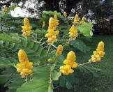
Antimicrobial Effect of Cassia alataLeaf Extracts on Fungal Isolates from Tinea Infections http://www.doi.org/10.26538/tjnpr/v7i5.29
Main Article Content
Abstract
Dermatophytoses are caused by fungi in the genera Microsprum, Trichophyton and Epidermophyton. Africans use extracts of medicinal plants to treat dermatophytosis. This study aimed to determine the in vitro antifungal activity of crude leaf extract of Cassia alata on fungal isolates from dermatophytosis lesions. Subjects with suspected lesions of dermatophytosis were recruited for the study. Skin scrapings were obtained from lesions for microscopy and culture. Isolates were identified macroscopically and microscopically. Fresh Cassia alata leaves were plucked and authenticated by a Botanist. Aqueous and alcohol extraction was done using the Soxhlet extractor. Isolates were subjected to in vitro leave extracts antifungal testing using disc diffusion methods. A total of 50 subjects were recruited for the study comprising 28(56.0%) males and 22(44.0%) females with a male to female ratio of 2.5: 1. The prevalence of dermatophytes infection in the study was 14(28.0%). Trichophyton rubrum was the most encountered isolates (42.8%). Males 10(71.4%) were more infected than females 4(28.5%). The susceptibility rates of dermatophytes to anfungals range between 0 100% with 30(78.6%) susceptibility to Griseofulvin. The aqueous extract was more effective with susceptibility rates 33.3% - 83.3% than ethanolic extract 16.7% - 50.0%. The dermatophytes were more susceptible to the 50µg/mL aqueous extract with rates between 50% - 83.3% while the range for the 50µg/mL ethanolic extract was 25.0% - 50.0%. Trichophyton veruccosum and E. floccosum were resistant to extracts. Cassia alata leaf extracts had antifungal activities against dermatophytes.
Downloads
Article Details

This work is licensed under a Creative Commons Attribution-NonCommercial-NoDerivatives 4.0 International License.
References
Liu D, Coloe S, Baird R, Pedersen J. Application of PCR to the identification of dermatophyte fungi. J. Med. Micro. 2000; 49:493–497.
Onayemi O, Isezuo SA, Njoku CH. Prevalence of different skin conditions in an outpatients setting in North-Western Nigeria. Int. J. Dermatol.2005; 44:7-11
Guerrant R, Walker D, Weller P. Tropical Infectious Diseases: Principles, Pathogens and Practice (3rd Edition). Philadelphia, Pa, USA: Elsevier Churchill Livingstone; 2011
Ayaya SO, Kamar KK, Kakai R. Aetiology of tinea capitis. East Afr. Med. J. 2000; 78:531–535.
Emele EF, Oyeka AC, Chika UF. Ringwm Infection in Nigeria: Investigating the Role of Barbers in Disease Transmission. Int. J. Public Health Res. 2015; III(2): 67-71.
Hennebelle T, Weniger B, Joseph H, Sahpaz S, Bailleul F. Senna alata. Fitoterapia 2009; 80(7): 385–393.
Manaharan T, Thirugnanasampandan R, Jayakumar R, Ramya G, Ramnath G, Kanthimathi MS. "Antimetastatic and Anti-Inflammatory Potentials of Essential Oil from Edible Ocimum sanctum Leaves". Sci. World J. 2014; 239508. https://doi.org/10.1155/2014/239508
Fatmawati S, Yuliana ASP, Abu Bakar, MF. Chemical constituents, usage and pharmacological activity of Cassia alata. Heliyon 2020; 6(7):e04396.
Oladeji OS, Adelowo FE, Oluyori AP, Bankole DT.Ethnobotanical Description and Biological Activities of Senna alata. Evid Based Complement Alternat Med. 2020; 2580259. doi: 10.1155/2020/2580259. eCollection 2020.
Adebayo O, Anderson WA, Moo-Young M, Snieckus V, Patil PA, Kolawole DO. “Phytochemistry and antibacterial activity of Senna alata flower.” PharmBiol.2001; 39(6): 408–412.
El-Mahmood AM, Doughari JH, Chanji FJ. “In vitro antibacterial activities of crude extract of Nauclealatifolia and Daniella oliver.” J. Sci. Res. Essay 2003; 31(3): 102– 105.
Oguntoye SO, Owoyale JA, Olatunji GA. “Antifungal and antibacterial activities of an alcoholic extract of S. alata leaves.”J. Appl. SCI. Environ. Manag. 2005; 9(3): 105–107.
Benjamin TV, Lamikanra A. “Investigation of Cassia alata, a plant used in Nigeria in the treatment of skin diseases.” Int.J.Crude Drug Res. 2008; 19(2-3): 93–96.
Leung L, Riutta T, Kotecha J, Rosser W. “Chronic constipation: an evidence-based review.” J Am Board Fam Med.2011; 24(4): 436–451.
Idu M, Omonigho SE, Igeleke CL. Preliminary investigation on the phytochemistry and antimicrobial activity of Senna alata L. flower. Pak. J. Biol. Sci.2007; 10: 806-809.
Dogo J, Afegbua LS, Dung CE. Prevalence of tinea capitis among School Children in Nok Community of Kaduna State, Nigeria. J Pathogens, 2016; 1-7.
Ogba OM, Bassey DE, Olorode OA, Mbah M. Barbing Equipments: Tools for Transmission of Tinea capitis. Microbiol. Res. J. Int. 2017; 19(1): 1-7, Article no.MRJI.31091
Ogba OM. Nyambi ES, Olorode OA, Enosakhare A. Antibacterial Activity of Lantana camara montevidensis Leaf Extract on Wound Isolates Using Animal Models. Trop J Nat Prod Res. 2021; 5(12):2160-2164.
Rex JH, Pfaller MA, Walsh TJ, Chaturvedi V, EspinelIngroff A, Ghannoum MA, Gosey LL, Odds FC, Rinaldi MG, Sheehan DJ, Warnock DW. Antifungal Susceptibility Testing: Practical Aspects and Current Challenges. Clin. Microbiol. Rev. 2006; 14(4): 643-658.
Barreto FS, Sousa EO, Campos AR, Costa JGM, Rodrigues FFG. Antibacterial Activity of Lantana camara linn and montevidensis Brig Extract from Cariri-Ceara, Brazil. J Young Pharm. 2010; 2(1):42-44.
Clinical and Laboratory Standards Institute (CLSI). Method for antifungal disk diffusion susceptibility testing nondermatophyte filamentous fungi; approved standard M51-A;Clinical and Laboratory Standards Institute: Wayne, PA, USA. 2010.
Adefemi AS, Odeigab OL, Alabi KM. Prevalence of dermatophytosisamong primary school children in Oke-Oyi Community of Kwara State, Nigeria. J. Cin. Practice 2011; 14(1): 23-27.
Ezihe MI, Magaji S, Bala M, Ibrahim R. Prevalence of Dermatophytes Infections among Public Primary School Children inBauchi Metropolis, of Bauchi State, Nigeria. Int. J. Sci. Eng.2017; 8(8): 1268
Abubacker MN, Ramanathan R, Kumar TS. In vitro antifungal activity of Cassia alata Linn. Flower extract. Nat. Prod. Radiance 2008; 7: 6-9
Sule WF, Okonkwo IO, Omo-Ogun S, Nwanze JC, Ojezele MO, Ojezele OJ. Phytochemical properties and in vitro antifungal activity of Senna alata Linn. Crude stem bark extract. J. Med. Plants Res. 2011; 5(2): 176-183.
Makinde A, Igoli JO, TA’Ama L. “Antimicrobial activity of Cassia alata”. Afr. J. Biotechnol. 2007; 6(13): 1509-1510


