

Sub-Acute Toxicity Profile of Aqueous Leaf Extract of Pavetta crassipes (K. Schum) in Rats http://www.doi.org/10.26538/tjnpr/v7i5.25
Article Sidebar
Main Article Content
Abstract
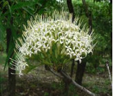
Pavetta crassipes is a plant with many ethnomedicinal uses but has limited published data on its safety. The aim of the study was to evaluate the subacute oral toxicity of aqueous leaf extract of P. crassipes in Wister rats. Thirty-six Wister male rats were randomly divided into 6 groups of six rats each. They were treated with; 1 mL/kg distilled water (negative control group), aqueous leaf extract of P. crassipes (125, 250 and 500 mg/kg test groups respectively), 10 mg/kg furosemide (positive control group 1), and 10 mg/kg lisinopril (positive control group 2) daily for 28 days. Weekly animal weight, food consumption, water intake, fecal/urine outputs, organ to
body weight ratio, hematological, biochemical and histological assessment of organs were used to evaluating toxicity. There was no significant difference between all the groups in body weight, food/water intake, fecal output, plasma sodium, potassium, chloride, calcium and biochemical markers of liver and kidney function (p›0.05). However, P. crassipes leaf extract significantly increases the urine excretion, high density lipoprotein (HDL) and endogenous antioxidant (p‹0.05) with a decrease in total cholesterol, triglyceride and low density lipoprotein (p‹0.05) in rats. Aqueous leaves extract of P. crassipes is relatively safe because the biochemical parameters and organs assayed were within the values for distilled water treated rats.
Downloads
Article Details

This work is licensed under a Creative Commons Attribution-NonCommercial-NoDerivatives 4.0 International License.
References
Bhusnure OG, Suryawanshi S, Vijayendra Swamy SM, Gholve SB, Girm PS, Birajdar MJ. Standardization and quality evaluation of herbal drugs. J. Drug Deliv. Ther.2019; 9(3):1058-1063
Radha KM, Puri S, Pundir A, Bangar SP, Changan S, Choudhary P, Parameswari E, Alhariri A, Samota MK, Damale RD, Singh S, Berwal MK, Dhumal S, Bhoite AG, Senapathy M, Sharma A, Bhushan B, Mekhemar M. Evaluation of nutritional, phytochemical, and mineral
composition of selected medicinal plants for therapeutic uses from cold desert of western Himalaya. Plants. 2021; 10(7):1429.doi.org/10.3390/plants10071429
van Wyk AS and Prinsloo G. Health, safety and quality concerns of plant-base traditional medicines and herbal remedies. S. Afr. J. Bot.2020;133:54-62.
Ibekwe NN, Adesomoju AA, Igoli JO, Barry III CE, Okogun JI. An iridoid glucoside from the leaves of Pavetta crassipes. J. Chem Soc. Nigeria.2019;44(4):629-632.
Patrick EB, Otimenyin SO, Bukar BB. Evaluation of Diuretic Potential of Aqueous Leaf Extract of Pavetta crassipes (K. Schum) in Rats. Trop J Nat Prod Res. 2022b; 6(5):801-805.
Ouattara LP, Sanon S, Mahiou-Leddet V, Gansané A, Baghdikian B, Traoré A, Nébié I,Traoré AS, Azas N, Ollivier E , Sirima SB. In vitro antiplasmodial activity of some medicinal plants of Burkina Faso. Parasitol. Res.2014; 113:405–416.
Bello IA, Ndukwe GI, Audu OT. Phytochemical analysis and biological activity of a precipitate from Pavetta crassipes.J. Med. Plants Res.2014;8:285-287.
Amos S, Okwuasaba FK, Gamaniel K, Akah P, Wambebe C. Inhibitory effects of the aqueous extract of Pavetta crassipes leaves on gastrointestinal and uterine smooth muscle preparations isolated from rabbits, guinea pigs and rats. JEthnopharmacology.1998; 61:209– 213.
Amos S, Akah PA, Binda L, Enwerem NM, Ogundaini A, Wambebe C, Hussaini IM, Gamaniel KS. Hypotensive activity of the ethanol extract of Pavetta crassipes leaves. Biol pharm bull. 2003;26(12):1674–1680. doi: 10.1248/bpb.26.1674.
Patrick EB, Otimenyin SO, Bukar BB. Survey of the blood pressure lowering potential of medicinal plants used in the management of hypertension in herbal homes in Zango Kataf, Kaduna, Nigeria. Nutr. Food Sci.. 2022a;53(1):178- 191. DOI 10.1108/NFS-11-2021-0326
Bariweni MW, Ozolua RI. Neuropharmacological effects of the aqueous leaf extract and fractions of Pavetta crassipes (K. Schum) Rubiaceae in mice. J Pharm Pharmacogn Res.2017; 5(5): 278–287.
Bariweni MW, Yibala OI, Ozolua RI.Toxicological studies on the aqueous leaf extract of Pavetta crassipes (K. Schum) in rodents. J Pharm Pharmacogn Res.2018; 6(1): 1–16
Committee for the Update of the Guide for the Care and Use of Laboratory Animals . Guide for the Care and Use of Laboratory Animals. 8th ed. National Academies Press; Washington, DC, USA: 2011
Ingle KP, Deshmukh AG, Padole DA, Dudhare MS, Moharil MP, Khelurkar VC. Phytochemicals: Extraction methods, identification, and detection of bioactive compounds from plant extracts. J Pharmacogn Phytochem. 2017; 6:32–36.
Barbara JB, Imelda B. Basic hematologic techniques. In: Lewis SM, Bain BJ, Bates I, (Eds.). Dacie and Lewis Practical Hematology. 11 th ed. London: Churchill Livingston; 2001.19-46 p.
Beutler E, Duron O, and Kelly BM. (1963). Improved Method for the Determination of Blood Glutathione. J. lab. clin. med.1963; 61: 882-888.
Rotruck JT, Pope AL, Ganther HE, Swanson AB, Hafeman DG, Hoekstra WG. Selenium: Biochemical Role as a Component of Glutathione Peroxidase. Science.1973; 179: 588-590. dx.doi.org/10.1126/science.179.4073.588
Claiborne A. Catalase activity. In: Greenwald, R.A., Ed., CRC Handbook of Methods for Oxygen Radical Research, CRC Press, Boca Raton, 1985;283-284.
Misra H, Fridovich I. The role of superoxide anion in the autoxidation of epinephrine to adrenochrome and a simple assay for superoxide dismutase. J. Biol. Chem.1972;247:3170–3175
Varshney R. and Kale RK. (1990). Effects of Calmodulin Antagonists on Radiation-Induced Lipid Peroxidation in Microsomes. Int. J. Radiat. Biol.1990; 58: 733-43. dx.doi.org/10.1080/09553009014552121
Galigher A E, Kozloff EN. Essentials of Practical Micro techniques. 2nd. Philadelphia, PA, USA: Lea & Febiger; 1971.
Mullens W, Damman K, Harjola V-P, Mebazaa A , BrunnetLa Rocca H-P, Martens P, Testani JM, Tang WHW, Or o F, Rossignol P, Metra M, Filippatos G, Seferovic PN, Ruschitzka F, Coats AL.The use of diuretics in heart failure with congestion — a position statement from the heart failure association of the European society of cardiology. Eur. J. Heart Fail.2019;21(2): 137-155
Lala V, Zubair M, Minter DA. Liver function tests. [online].2022[cited 2022 Feb 24]. Available from: https://www.ncbi.nlm.nih.gov/books/NBK482489/
Gounden V, Bhatt H, Jialal I. Renal function test. [online].2022 [cited 2022 Feb 24]. Available from: https://www.ncbi.nlm.nih.gov/books/NBK507821/
Wang Y, Branicky R, Noe A, Hekimi S.Superoxide dismutases:dual roles in controlling ROS damage and regulating ROS signaling. J Cell Biol. 2017;217(6):1915- 1928.
Ighodaro OM, and Akinloye OA. First line defense antioxidants-superoxide dismutase (SOD), catalase (CAT) and glutathione peroxidase (GPX): their fundamental role in the entire antioxidant defense grid. Alexandria J. Med.2018;54:287-293.
Nandi A, Yan L-J,Jana CK, Das N.Role of catalase in oxidative stress and age associated degenerative diseases. Oxid Med Cell Longev. 2019;2019:9613090. doi: 10.1155/2019/9613090
Andrés CMC, Pérez de la Lastra JM, Juan CA, Plou FJ, Pérez-Lebeña E. Chemistry of Hydrogen Peroxide Formation and Elimination in Mammalian Cells, and Its Role in Various Pathologies. Stresses. 2022; 2(3):256-274. doi.org/10.3390/stresses2030019
Behnisch-cornwell S, Wolff L, Bednarski PJ.The effect of glutathione peroxidase-1 knockout on anticancer sensitivities and reactive oxygen species in haploid HAP-1 cells. Antioxidants.2020; 9(12):1300. doi.org/10.3390/antiox9121300
Kwon DH, Cha H-J, Lee H, Hong S-H, Park C, Park S-H, Kim G-Y, Kim S, Kim H-S, Hwang H-J, Cho YH.Protective effect of glutathione against oxidative stress-induced cytotoxicity in RAW 264.7 macrophages through activating the nuclear factor erythroid 2- related factor-2/heme oxygenase-1 pathway. Antioxidants (Basel).2019;8(4): 82. . doi: 10.3390/antiox8040082
. Kumari S, Deori M, Elancheran R, Kotoky J and Devi R (2016) In vitro and In vivo Antioxidant, Anti-hyperlipidemic Properties and Chemical Characterization of Centella asiatica (L.) Extract. Front. Pharmacol. 7:400. doi: 10.3389/fphar.2016.00400
Wang, H., Gao, X. D., Zhou, G. C., Cai, L., and Yao, W. B. In vitro and in vivo antioxidant activity of aqueous extract from Choerospondias axillaris fruit. Food Chem. 2008; 106:888–895.
Aremu.O.O, Oyedeji, A.O, Oyedeji, O.O, Nkeh-Chungag,B.N, Rusike,C.R.S.(2019).Invitro and In vivo antioxidant properties of Taraxacum officinale in NѠ-Nitro-L-Arginine Methyl Ester(L-NAME)- induce hypertensive Rats. Antioxidants 2019; 8(8):309. doi.org/10.3390/antiox808030
Mas-Bargues C, Escrivá C, Dromant M, Borrás C, Viña J. Lipid peroxidation as measured by chromatographic determination of malondialdehyde. human plasma reference values in health and disease. Archives of biochemistry and biophysics Arch.Biochem. Bio.phys.2021;709:108941. doi.org/10.1016/j.abb.2021.108941